Hormone Replacement Therapy (HRT) in India — Is It Safe and Who Should Consider It?

Few medical topics have been more misunderstood, more feared, and more under-prescribed in India than hormone replacement therapy (HRT). A study published in 2002 sent women and their doctors into panic — and its flaws and subsequent corrections were never communicated with the same urgency as the original alarm. Two decades later, the science on HRT has been thoroughly re-evaluated. The result is reassuring: for most healthy women under 60 who begin HRT within 10 years of menopause, the benefits substantially outweigh the risks. Dr. Shweta Mendiratta, menopause specialist in Faridabad, provides a clear, evidence-based guide.

What Is HRT?

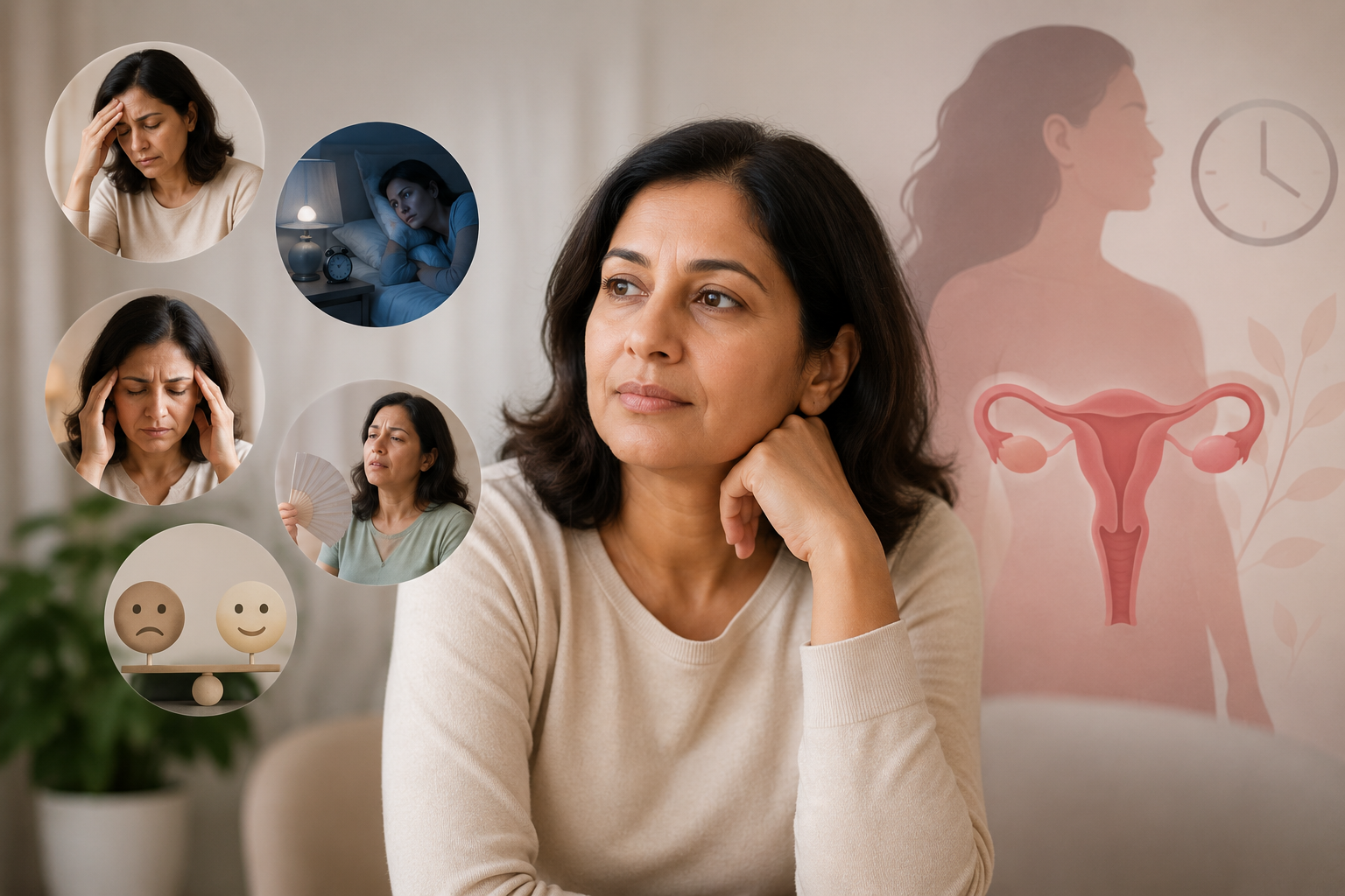
Hormone replacement therapy replaces the oestrogen (and where applicable, progesterone) that the ovaries stop producing at menopause. It is used to:

  • Relieve menopausal symptoms (hot flushes, night sweats, sleep disturbances, mood changes, vaginal dryness)
  • Protect bone density and reduce fracture risk
  • Reduce cardiovascular risk when started early in the menopause transition
  • Improve quality of life, cognitive function, and sexual health

Types of HRT Available in India

By Hormone Type

  • Oestrogen-only HRT: For women who have had a hysterectomy and have no uterus
  • Combined HRT (oestrogen + progestogen): For women with an intact uterus; the progestogen protects the uterine lining from the effects of unopposed oestrogen
  • Body-identical HRT: Uses micronised progesterone (identical in structure to natural progesterone) combined with oestradiol — associated with a lower risk profile than older synthetic progestogens

By Route of Administration

  • Oral tablets: Convenient but undergo first-pass metabolism in the liver
  • Transdermal patches: Applied to the skin twice weekly; bypass liver metabolism; lower risk of blood clots
  • Gels: Oestrogen gel applied daily to the skin; excellent alternative for women who dislike patches
  • Vaginal oestrogen (cream, pessary, ring): For local genitourinary symptoms only; negligible systemic absorption

Transdermal and vaginal oestrogen routes are increasingly preferred because they avoid first-pass hepatic metabolism and carry a lower risk of venous thromboembolism (blood clots) compared to oral oestrogen.

The Benefits of HRT — What the Evidence Shows

Symptom Relief

HRT is the most effective treatment for vasomotor symptoms (hot flushes and night sweats), with reduction rates of 75–90%. It is also highly effective for vaginal dryness, sleep disruption, mood changes, and joint pain associated with menopause.

Bone Protection

Oestrogen is the primary regulator of bone turnover in women. HRT reduces the rate of bone loss, prevents osteoporosis, and significantly reduces the risk of hip and vertebral fractures. It is particularly important for women who reach menopause before age 45.

Cardiovascular Health

When started within 10 years of menopause (the ‘window of opportunity’), oestrogen has beneficial effects on the cardiovascular system — improving lipid profiles, reducing arterial stiffness, and potentially reducing the risk of coronary heart disease. Women who begin HRT close to menopause appear to have a reduced risk of cardiovascular events compared to women who delay or never use HRT.

Cognitive Function

Oestrogen plays a role in brain function and neuroprotection. Emerging evidence suggests that HRT started early in the menopause transition may reduce the risk of Alzheimer’s disease, though this remains an area of active research.

The Risks of HRT — Understanding the Real Numbers

Breast Cancer

The most feared risk associated with HRT is breast cancer. The picture is nuanced:

  • Oestrogen-only HRT (for women without a uterus) is not associated with a significantly increased breast cancer risk and may even be associated with a slight reduction in risk
  • Combined HRT (oestrogen + synthetic progestogen) is associated with a small increased risk — approximately 4 extra cases per 1000 women over 5 years of use
  • Body-identical HRT using micronised progesterone appears to carry a lower breast cancer risk than synthetic progestogen combinations
  • The excess risk is comparable to that from drinking one to two glasses of alcohol per day or being overweight
  • Breast cancer risk returns to baseline within 5 years of stopping HRT

Blood Clots (VTE)

Oral oestrogen doubles the baseline risk of venous thromboembolism (DVT/PE). However, transdermal oestrogen does not increase VTE risk — a crucial distinction. For women at increased VTE risk, transdermal HRT is the preferred route.

Stroke

Oral oestrogen is associated with a small increase in ischaemic stroke risk; transdermal oestrogen is not. Again, route of administration matters significantly.

Who Should Consider HRT?

HRT is appropriate for:

  • Women with significant menopausal symptoms affecting quality of life
  • Women with premature ovarian insufficiency (POI) or early menopause — HRT is essential for bone and cardiovascular protection and should be continued until the natural age of menopause
  • Women with osteoporosis or high fracture risk
  • Women who have failed or prefer not to use non-hormonal alternatives for symptom management

Who Should Not Use HRT?

  • Women with current or recent hormone receptor-positive breast cancer (oestrogen-sensitive)
  • Women with unexplained vaginal bleeding
  • Women with active thromboembolism (DVT or PE)
  • Women with severe liver disease

Note that a history of cardiovascular disease, migraine with aura, or controlled hypertension is not an absolute contraindication to HRT — the route, type, and dose can be adjusted. Dr. Mendiratta conducts a thorough risk assessment before prescribing.

HRT in India: Availability and Cost

Several HRT preparations are available in India, including oral oestradiol and combined preparations. Transdermal patches and gels are increasingly available in urban pharmacies, though access varies. Body-identical micronised progesterone is available. Dr. Mendiratta can advise on the most appropriate formulation for your needs and budget.

Frequently Asked Questions (FAQs)

Q1. How long can I take HRT?

There is no arbitrary maximum duration. HRT should be used for as long as you need it and the benefits outweigh the risks for you individually. An annual review with your doctor is recommended. Many women use HRT safely for 5–10 years or longer.

Q2. Will I gain weight on HRT?

Menopause itself causes weight gain — particularly abdominal fat — due to falling oestrogen. HRT may actually mitigate this effect. Clinical studies do not consistently show that HRT causes weight gain beyond what is expected in the menopause transition.

Q3. Can I start HRT 10 years after menopause?

Starting HRT more than 10 years after menopause or after age 60 is more complex. The cardiovascular and cognitive benefits are less certain and there may be a slightly higher risk of cardiovascular events. This needs to be assessed individually.

Q4. Is HRT available over the counter in India?

HRT is a prescription medication in India and should only be initiated under medical supervision after a full assessment of your health history, risk factors, and symptom profile.

Q5. What is bio-identical HRT and is it better?

‘Bio-identical’ refers to hormones that are structurally identical to those produced by the body (oestradiol and micronised progesterone). These are available as regulated pharmaceutical preparations and are generally preferred over compounded alternatives. They appear to carry a more favourable safety profile, particularly regarding breast cancer risk, than older synthetic progestogens.


Get In Touch With Dr. Shweta Mendiratta

Phone: +91-8130048652 | +91-9999093503

Email: shwetasmendiratta@gmail.com

Yatharth Super Speciality Hospital

Plot No 9, Sector-20, Krishna Nagar, New Industrial Township, Faridabad, Haryana 121007

Phone: +91 8178-939442

Mediclub Gynae ‘N’ Neuro Clinic

Plot No. 857 Sector 21 C, Faridabad Delhi, Haryana 121001

Perimenopause at 40 — Signs Most Indian Doctors Miss and What to Do

Most women expect menopause to arrive somewhere in their early to mid-fifties. So when changes begin in their late thirties or early forties — irregular periods, sleepless nights, inexplicable mood swings, brain fog — they rarely connect them to hormonal transition. Neither, unfortunately, do many doctors, who dismiss these symptoms as stress, anxiety, or thyroid problems without considering the most likely culprit: perimenopause. Dr. Shweta Mendiratta, menopause specialist in Faridabad, explains what perimenopause is, why it is so often missed, and what women can do about it.

What Is Perimenopause?

Perimenopause is the transitional phase leading up to menopause — the permanent cessation of menstruation. It typically begins 4–8 years before the last menstrual period. For most women, this means perimenopause starts between ages 40 and 51, though it can begin as early as the late thirties.

Menopause itself is defined as 12 consecutive months without a period. Perimenopause is everything before that point. During this phase, the ovaries gradually produce less oestrogen and progesterone, and ovulation becomes irregular.

Why Perimenopause Is Missed in India

Several factors contribute to the underdiagnosis of perimenopause in Indian women:

  • Cultural normalisation of suffering: Mood changes, fatigue, and physical discomfort are often attributed to work stress, family responsibilities, or simply ‘getting older.’
  • Lack of awareness: Perimenopause is rarely discussed openly in Indian families or medical consultations.
  • Symptom overlap: Perimenopause symptoms mimic thyroid disease, depression, anxiety, anaemia, and other common conditions.
  • Diagnostic bias: Doctors often treat the individual symptoms (sleeping tablets for insomnia, antidepressants for mood changes) without considering the underlying hormonal cause.

Symptoms of Perimenopause — The Complete Picture

Menstrual Changes

  • Irregular periods — cycles becoming shorter (less than 21 days) or longer (more than 35 days)
  • Heavier or lighter than usual periods
  • Missed periods — often mistaken for pregnancy
  • Intermenstrual spotting

Vasomotor Symptoms

  • Hot flushes: Sudden waves of heat, typically in the face, neck, and chest, often accompanied by sweating
  • Night sweats: Drenching sweats during sleep, leading to disturbed sleep and chronic fatigue

Sleep Disturbances

Many perimenopausal women develop insomnia — difficulty falling asleep, frequent waking, or early morning waking — that has no other apparent cause. This is directly linked to falling oestrogen and progesterone levels.

Mood and Cognitive Changes

  • Increased anxiety, irritability, or emotional lability
  • Low mood or mild depression
  • Brain fog: Difficulty concentrating, poor short-term memory, word-finding difficulties

These symptoms are frequently misattributed to clinical depression or anxiety disorder and treated with antidepressants or anxiolytics when hormone therapy would be more appropriate.

Physical Symptoms

  • Vaginal dryness and reduced lubrication (genitourinary syndrome of menopause)
  • Reduced libido
  • Joint and muscle aches
  • Headaches
  • Changes in skin (increased dryness, loss of elasticity)
  • Weight gain, particularly around the abdomen

How Is Perimenopause Diagnosed?

Perimenopause is primarily a clinical diagnosis based on age, symptoms, and menstrual history. Blood tests can be supportive but are not always definitive, particularly in early perimenopause when hormone levels fluctuate widely:

  • FSH: Rising FSH (above 10 IU/L, particularly above 25 IU/L) suggests reduced ovarian reserve
  • Oestradiol: Fluctuating, often lower than expected
  • AMH: Declining; a very low AMH in a symptomatic woman in her 40s is consistent with perimenopause
  • Thyroid function: Essential to rule out as a contributing factor
  • Full blood count: To exclude anaemia as a cause of fatigue

What Women Can Do

Lifestyle Measures

  • Regular strength training and weight-bearing exercise (to protect bone density and muscle mass)
  • Aerobic exercise (to improve cardiovascular health and mood)
  • Reducing alcohol and caffeine (which worsen hot flushes and sleep disruption)
  • Adequate calcium and vitamin D intake
  • Stress management through yoga, mindfulness, or counselling

Hormone Replacement Therapy (HRT)

For women with significant perimenopausal symptoms, HRT is the most effective treatment. Modern HRT — using body-identical hormones at the lowest effective dose — is safe for most women under 60 who begin it within 10 years of menopause. Dr. Mendiratta provides individualised HRT assessment and prescribing, including discussion of risks and benefits in the context of your personal health history.

Non-Hormonal Options

For women who cannot or prefer not to use HRT, several non-hormonal options help manage specific symptoms:

  • SSRIs/SNRIs: For hot flushes and mood symptoms
  • Clonidine: For hot flushes
  • Cognitive behavioural therapy (CBT): Highly effective for mood and sleep disturbances
  • Vaginal oestrogen: Safe even for women who cannot use systemic HRT; treats genitourinary symptoms locally

Frequently Asked Questions (FAQs)

Q1. Can I get pregnant during perimenopause?

Yes. Ovulation still occurs intermittently during perimenopause, and pregnancy is possible. Contraception should be continued until you have had 12 consecutive months without a period (i.e., confirmed menopause) or until age 55.

Q2. What is the difference between perimenopause and premature ovarian insufficiency (POI)?

POI (previously called premature menopause) occurs before age 40. Perimenopause is a natural transition that begins in the 40s. Both are characterised by declining ovarian function, but POI is less expected and has different implications for fertility and bone health.

Q3. How long does perimenopause last?

On average, 4–8 years, but it can be shorter or longer. Symptoms may be most intense in the 1–2 years just before the last period.

Q4. Is it normal to have periods every 2 weeks during perimenopause?

Irregular cycles, including short cycles, are common in perimenopause. However, very frequent or heavy bleeding should be evaluated to rule out fibroids, polyps, or endometrial abnormalities.

Q5. Does perimenopause affect bone health?

Yes. The decline in oestrogen during perimenopause accelerates bone loss. This is a key reason why calcium, vitamin D, weight-bearing exercise, and HRT (when appropriate) are important during this phase.


Get In Touch With Dr. Shweta Mendiratta

Phone: +91-8130048652 | +91-9999093503

Email: shwetasmendiratta@gmail.com

Yatharth Super Speciality Hospital

Plot No 9, Sector-20, Krishna Nagar, New Industrial Township, Faridabad, Haryana 121007

Phone: +91 8178-939442

Mediclub Gynae ‘N’ Neuro Clinic

Plot No. 857 Sector 21 C, Faridabad Delhi, Haryana 121001

Preeclampsia — The Silent Pregnancy Danger Every Indian Family Must Know About

Preeclampsia is responsible for approximately 17% of maternal deaths in India — making it one of the most dangerous complications of pregnancy and one that every pregnant woman, her family, and her doctor must take seriously. Yet it often develops with no obvious warning signs, which is why it is called the silent danger of pregnancy. Dr. Shweta Mendiratta, high-risk obstetrics specialist in Faridabad, explains what preeclampsia is, who is at risk, and what every family must know.

What Is Preeclampsia?

Preeclampsia is a serious pregnancy complication characterised by high blood pressure (hypertension) that develops after 20 weeks of gestation in a woman who previously had normal blood pressure, combined with signs of damage to organ systems — most often the kidneys (manifesting as protein in the urine), liver, brain, or placenta.

Without timely diagnosis and management, preeclampsia can rapidly progress to eclampsia (seizures), HELLP syndrome (a life-threatening combination of haemolysis, elevated liver enzymes, and low platelet count), stroke, kidney failure, and maternal or fetal death.

Who Is at Risk?

  • First-time pregnancies (nulliparity)
  • Pre-existing hypertension or kidney disease
  • Diabetes (type 1, type 2, or gestational)
  • Multiple pregnancy (twins, triplets)
  • Obesity (BMI above 30)
  • Family history of preeclampsia
  • Previous preeclampsia in a prior pregnancy
  • Autoimmune conditions (lupus, antiphospholipid syndrome)
  • Age over 40
  • Short interval between pregnancies (less than 2 years) or very long interval (over 10 years)

Recognising the Warning Signs

Many women with preeclampsia have no symptoms — which is why regular blood pressure monitoring at every antenatal visit is non-negotiable. However, warning signs that require immediate medical attention include:

  • Sudden or severe headache not relieved by paracetamol
  • Visual disturbances: blurring, flashing lights, or temporary vision loss
  • Severe upper abdominal or right-sided pain (a sign of liver involvement)
  • Sudden swelling of the face, hands, or feet (especially if rapid and severe)
  • Feeling very unwell or confused
  • Decreased fetal movements

If you experience any of these symptoms in pregnancy, go to the hospital immediately — do not wait for your next routine appointment.

How Is Preeclampsia Diagnosed?

Blood pressure: Readings of 140/90 mmHg or above on two occasions at least 4 hours apart (or 160/110 mmHg on a single reading) after 20 weeks of gestation.

Urine testing: Proteinuria (protein in the urine) detected by dipstick or confirmed by a 24-hour urine collection.

Blood tests: Full blood count (platelets), liver enzymes (ALT, AST), creatinine, uric acid, and LDH — to assess organ involvement.

Fetal monitoring: Ultrasound for fetal growth, amniotic fluid, and Doppler studies of umbilical blood flow — preeclampsia can restrict placental blood flow and impair fetal growth.

Management of Preeclampsia

Mild to Moderate Preeclampsia

For mild preeclampsia, close monitoring may allow the pregnancy to continue with:

  • Antihypertensive medications to keep blood pressure below 150/100 mmHg
  • Regular blood tests and fetal monitoring
  • Rest, though strict bed rest is no longer recommended
  • Low-dose aspirin (started before 16 weeks in high-risk women) for prevention

Severe Preeclampsia

Severe preeclampsia (BP above 160/110, or with severe organ involvement) typically requires hospital admission, IV antihypertensive therapy, intravenous magnesium sulphate (to prevent seizures), and plans for delivery as soon as it is safe.

Delivery

The only definitive cure for preeclampsia is delivery. The timing of delivery balances the risks to the mother of continuing the pregnancy against the risks to the baby of preterm birth. Dr. Mendiratta works with the neonatology team to optimise the timing and mode of delivery for each patient.

Preeclampsia Prevention

For women identified as high risk, daily low-dose aspirin (75–150 mg) started before 16 weeks of gestation has strong evidence for reducing the risk of preeclampsia. Calcium supplementation (1.5–2g per day) is also recommended in women with low dietary calcium intake, which is common in India.

Frequently Asked Questions (FAQs)

Q1. Can preeclampsia recur in a subsequent pregnancy?

Yes. Women who have had preeclampsia have a 20–25% risk of it recurring in future pregnancies. This risk is higher if the preeclampsia was severe or early-onset. Close monitoring and prophylactic aspirin in subsequent pregnancies are essential.

Q2. Does preeclampsia affect the baby?

Preeclampsia restricts blood flow through the placenta, which can impair fetal growth (intrauterine growth restriction). If delivery is required prematurely, the baby may need neonatal intensive care.

Q3. Will preeclampsia go away after delivery?

In most cases, blood pressure and urine protein normalise within 6–12 weeks after delivery. However, women who have had preeclampsia have a higher lifelong risk of hypertension, stroke, and cardiovascular disease and should be monitored accordingly.

Q4. Is home blood pressure monitoring safe in pregnancy?

Yes, and it is recommended for women at risk of preeclampsia. Regular home monitoring allows earlier detection of rising blood pressure between clinic visits.

Q5. Can I have preeclampsia without protein in my urine?

Yes. Revised criteria for preeclampsia no longer require proteinuria if there is evidence of other organ involvement — such as low platelets, elevated liver enzymes, impaired kidney function, or neurological symptoms.


Get In Touch With Dr. Shweta Mendiratta

Phone: +91-8130048652 | +91-9999093503

Email: shwetasmendiratta@gmail.com

Yatharth Super Speciality Hospital

Plot No 9, Sector-20, Krishna Nagar, New Industrial Township, Faridabad, Haryana 121007

Phone: +91 8178-939442

Mediclub Gynae ‘N’ Neuro Clinic

Plot No. 857 Sector 21 C, Faridabad Delhi, Haryana 121001

Gestational Diabetes in Faridabad — Managing Sugar in Pregnancy Step by Step

Gestational diabetes mellitus (GDM) is one of the most common complications of pregnancy, affecting 10–20% of pregnant women in India — significantly higher than the global average. Yet it is also one of the most manageable conditions when identified early and addressed systematically. Dr. Shweta Mendiratta, obstetrician and high-risk pregnancy specialist in Faridabad, walks you through everything you need to know about gestational diabetes — from what causes it to how it is managed to what happens after delivery.

What Is Gestational Diabetes?

Gestational diabetes is a form of glucose intolerance that develops during pregnancy in women who did not have diabetes before. During pregnancy, the placenta produces hormones that can cause insulin resistance — particularly in the second and third trimesters. In women who cannot produce enough extra insulin to compensate, blood sugar levels rise, resulting in GDM.

GDM differs from pre-existing diabetes (type 1 or type 2), though the distinction between GDM and undiagnosed pre-existing diabetes discovered in pregnancy is sometimes difficult.

Why Is GDM More Common in India?

Indians are genetically predisposed to insulin resistance at lower body weights than Caucasians. The ‘Asian phenotype’ — relatively higher body fat percentage at a given BMI, a predisposition to abdominal fat, and a high-carbohydrate traditional diet — creates the perfect storm for gestational diabetes. Rapid urbanisation, increasingly sedentary lifestyles, and rising rates of obesity amplify this risk further.

Who Is at Risk?

  • Pre-pregnancy BMI above 23 kg/m² (lower threshold for Asians)
  • Family history of type 2 diabetes
  • Previous gestational diabetes
  • Previous macrosomic baby (birth weight above 3.5–4 kg)
  • PCOS
  • Age above 35
  • Multiple pregnancy (twins or more)

Screening and Diagnosis

In India, universal screening for GDM is recommended for all pregnant women. Dr. Mendiratta follows the DIPSI (Diabetes in Pregnancy Study Group India) or IADPSG protocol, depending on your specific situation:

  • 75g oral glucose tolerance test (OGTT) between 24–28 weeks of gestation — or earlier if risk factors are present
  • Diagnosis is confirmed if fasting glucose is ≥92 mg/dL, or 1-hour post-glucose is ≥180 mg/dL, or 2-hour post-glucose is ≥153 mg/dL

What Happens If GDM Is Not Treated?

Risks to the Baby

  • Macrosomia (excessive birth weight), leading to difficult delivery or caesarean section
  • Shoulder dystocia (dangerous delivery complication)
  • Neonatal hypoglycaemia (low blood sugar after birth)
  • Respiratory distress
  • Higher risk of obesity and type 2 diabetes in childhood

Risks to the Mother

  • Preeclampsia
  • Higher rate of caesarean delivery
  • Up to 50% risk of developing type 2 diabetes within 5–10 years after delivery

Step-by-Step Management of GDM

Step 1: Medical Nutrition Therapy

Diet is the cornerstone of GDM management. Dr. Mendiratta works with patients to develop an individualised diet plan:

  • Distribute carbohydrates evenly across 3 main meals and 2–3 snacks
  • Choose complex, low-GI carbohydrates (millets, oats, legumes, vegetables)
  • Limit refined grains, sweets, fruit juices, and sugary beverages
  • Include adequate protein and healthy fats at every meal
  • Monitor portion sizes — do not skip meals

Step 2: Physical Activity

Regular moderate exercise improves insulin sensitivity. Walking for 30 minutes after meals is particularly effective at blunting post-meal glucose spikes. Pregnancy-safe exercise, including walking, yoga, and swimming, is encouraged unless medically contraindicated.

Step 3: Blood Glucose Monitoring

Self-monitoring of blood glucose (SMBG) is recommended:

  • Fasting glucose target: Below 95 mg/dL
  • 1-hour post-meal target: Below 140 mg/dL
  • 2-hour post-meal target: Below 120 mg/dL

Step 4: Insulin or Oral Medication

If blood glucose targets are not met within 1–2 weeks of diet and exercise changes, medication is required. Insulin is the safest and most effective option in pregnancy. Metformin may be used in selected cases. Dr. Mendiratta will titrate insulin doses carefully based on your glucose log.

Step 5: Fetal Monitoring

Women with GDM require additional monitoring including regular growth scans, Doppler studies of umbilical blood flow, and non-stress tests — particularly in the third trimester.

After Delivery

Blood glucose levels typically normalise after delivery, but GDM leaves a lasting metabolic imprint. Dr. Mendiratta recommends a 75g OGTT at 6–12 weeks postpartum to confirm normoglycaemia, followed by annual fasting glucose checks. Breastfeeding, maintaining a healthy weight, and regular exercise significantly reduce the risk of future type 2 diabetes.

Frequently Asked Questions (FAQs)

Q1. Does GDM mean I will have diabetes for life?

No. GDM typically resolves after delivery. However, it is a significant risk factor for type 2 diabetes in the future — approximately 50% of women with GDM develop type 2 diabetes within 5–10 years. Lifestyle modification dramatically reduces this risk.

Q2. Can I have a normal vaginal delivery with GDM?

Yes, in most cases. The decision on mode of delivery depends on your glucose control, the baby’s estimated weight, and other obstetric factors.

Q3. Is it safe to take insulin during pregnancy?

Insulin does not cross the placenta and is completely safe for the baby. It is actually the safest and most predictable treatment for GDM.

Q4. What should I eat for breakfast if I have GDM?

A GDM-friendly breakfast includes a small portion of protein (eggs, paneer, dahi), healthy fat (nuts), and a small amount of complex carbohydrate (1 small roti, oats, or millet). Avoid fruit juice, white bread, and sugary cereals in the morning, when insulin resistance is typically highest.

Q5. Can GDM be prevented?

Preconception weight management, a low-GI diet, and regular physical activity can reduce the risk but cannot guarantee prevention in genetically predisposed women.


Get In Touch With Dr. Shweta Mendiratta

Phone: +91-8130048652 | +91-9999093503

Email: shwetasmendiratta@gmail.com

Yatharth Super Speciality Hospital

Plot No 9, Sector-20, Krishna Nagar, New Industrial Township, Faridabad, Haryana 121007

Phone: +91 8178-939442

Mediclub Gynae ‘N’ Neuro Clinic

Plot No. 857 Sector 21 C, Faridabad Delhi, Haryana 121001


Ovarian Cystectomy — When to Watch and When to Operate, Explained by Dr. Shweta Mendiratta

An ovarian cyst diagnosis can be alarming — but in most cases, the cyst is benign, causes no symptoms, and resolves on its own. The challenge is identifying which cysts require surveillance only, which need medical management, and which genuinely need surgical removal. Dr. Shweta Mendiratta, laparoscopic and robotic gynaecological surgeon in Faridabad, guides you through the decision-making process.

What Are Ovarian Cysts?

An ovarian cyst is a fluid-filled sac that develops on or within an ovary. They are extraordinarily common — most women will have at least one ovarian cyst during their lifetime, often without knowing it.

There are several types of ovarian cysts:

  • Functional cysts: Follicular cysts (from an egg-containing follicle that doesn’t rupture) and corpus luteum cysts (from the structure left after ovulation). These are by far the most common and almost always resolve within 1–3 menstrual cycles without treatment.
  • Endometriomas (chocolate cysts): Cysts caused by endometriosis, filled with old blood. These do not resolve spontaneously and can impair ovarian reserve.
  • Dermoid cysts (mature teratomas): Contain a variety of tissue types (skin, hair, teeth). Generally benign but require surgical removal due to the risk of complications.
  • Cystadenomas: Serous or mucinous cysts arising from the ovarian surface. Can grow large; require surgical removal.
  • Polycystic ovaries: Multiple small follicles in PCOS — not ‘cysts’ in the traditional sense.

When to Watch and Wait

Not all ovarian cysts require surgery. Expectant management (watching and waiting) is appropriate when:

  • The cyst is less than 5 cm in a premenopausal woman
  • The ultrasound features are reassuringly simple (thin-walled, no solid components, no septae, no blood flow within the cyst)
  • The woman is asymptomatic
  • Tumour markers (CA-125, AFP, inhibin) are normal
  • The cyst is consistent with a functional cyst

In these cases, Dr. Mendiratta typically recommends a repeat ultrasound after 6–12 weeks to confirm resolution.

When Surgery Is Required

Surgical removal (ovarian cystectomy) is recommended when:

  • The cyst is large (generally over 5–6 cm and not decreasing in size)
  • Ultrasound features are suspicious: thick walls, internal septations, solid components, papillary projections, or abnormal blood flow
  • CA-125 or other tumour markers are elevated
  • The cyst persists beyond 3 menstrual cycles
  • The cyst is causing significant pain, pressure, or other symptoms
  • Torsion (twisting of the ovary) is suspected — a gynaecological emergency
  • The cyst has ruptured and is causing internal bleeding
  • The woman is postmenopausal (any new cyst requires careful evaluation)
  • The cyst is an endometrioma (particularly if affecting fertility or growing)
  • The cyst is a dermoid or cystadenoma (which do not resolve spontaneously)

Ovarian Cystectomy: The Surgical Procedure

Ovarian cystectomy — removal of the cyst while preserving the ovary — is the preferred approach for most benign cysts in women of reproductive age. Dr. Mendiratta performs this laparoscopically or robotically:

  • 3–4 small port incisions (5–10 mm)
  • The cyst wall is carefully dissected from the ovarian tissue and removed intact
  • Ovarian tissue is meticulously preserved
  • The ovary is repaired (no sutures needed in small defects; careful suturing for larger ones)

Preservation of healthy ovarian tissue is critically important, particularly for women who wish to conceive. Dr. Mendiratta uses technique specifically designed to minimise damage to the surrounding follicles.

Endometrioma Surgery — Special Considerations

Ovarian endometriomas (chocolate cysts) require particularly careful surgical technique because the cyst wall is adherent to normal ovarian cortex and indiscriminate removal damages the healthy tissue beneath. Evidence suggests that stripping endometriomas significantly reduces ovarian reserve (AMH levels). Dr. Mendiratta uses a conservative, minimal-damage technique and discusses the risks and benefits of surgery versus expectant management thoroughly with patients planning IVF.

Frequently Asked Questions (FAQs)

Q1. Can ovarian cysts cause infertility?

Functional cysts do not cause infertility and resolve on their own. Endometriomas and large cysts that damage ovarian tissue can impair fertility. The decision to operate should carefully weigh the potential damage from surgery against the damage from leaving the cyst in place.

Q2. Is ovarian cyst surgery safe?

Laparoscopic ovarian cystectomy is a safe, well-established procedure. As with all surgery, there are small risks of bleeding, infection, and injury to adjacent structures, which are minimised by the experience of the surgeon.

Q3. How quickly do ovarian cysts regrow after surgery?

Simple functional cysts do not recur. Endometriomas have a recurrence rate of approximately 20–30% over 5 years. Medical treatment (OCPs, dienogest) after surgery reduces recurrence risk.

Q4. Can an ovarian cyst be cancerous?

The vast majority of ovarian cysts in women under 50 are benign. However, certain ultrasound features (solid components, internal blood flow, papillary projections) and elevated tumour markers raise the suspicion of malignancy and require urgent specialist evaluation.

Q5. My doctor said my cyst is 4 cm — should I be worried?

A 4 cm cyst in a premenopausal woman with benign ultrasound features is unlikely to be serious. A repeat scan in 6–8 weeks is typically advised. If it persists or grows, further evaluation is warranted.


Get In Touch With Dr. Shweta Mendiratta

Phone: +91-8130048652 | +91-9999093503

Email: shwetasmendiratta@gmail.com

Yatharth Super Speciality Hospital

Plot No 9, Sector-20, Krishna Nagar, New Industrial Township, Faridabad, Haryana 121007

Phone: +91 8178-939442

Mediclub Gynae ‘N’ Neuro Clinic

Plot No. 857 Sector 21 C, Faridabad Delhi, Haryana 121001

Robotic Myomectomy for Fibroids — Preserving the Uterus Without Open Surgery

Uterine fibroids are extraordinarily common — they affect up to 70% of women by age 50, though not all cause symptoms. For women who have problematic fibroids but wish to preserve their uterus — whether for future pregnancy, personal preference, or cultural reasons — myomectomy (surgical removal of fibroids while keeping the uterus intact) is the answer. And today, thanks to robotic and laparoscopic technology, myomectomy can be performed with minimal incisions, minimal blood loss, and a rapid return to normal life. Dr. Shweta Mendiratta, robotic surgeon in Faridabad, explains everything you need to know.

What Are Uterine Fibroids?

Fibroids (leiomyomas or myomas) are non-cancerous growths arising from the muscle tissue of the uterus. They vary enormously in size — from a few millimetres to several centimetres — and in location:

  • Intramural: Within the uterine muscle wall (most common)
  • Submucosal: Projecting into the uterine cavity (most likely to cause heavy bleeding and fertility problems)
  • Subserosal: Projecting outward from the uterine surface
  • Pedunculated: Attached to the uterus by a stalk

A woman may have a single fibroid or many (multiple fibroids), and the distribution matters for surgical planning.

When Do Fibroids Need Treatment?

Many fibroids are asymptomatic and require no treatment beyond monitoring. Treatment is recommended when fibroids cause:

  • Heavy or prolonged menstrual bleeding (leading to anaemia)
  • Pelvic pain or pressure
  • Urinary frequency or difficulty emptying the bladder
  • Constipation or rectal pressure
  • Difficulty conceiving or recurrent miscarriage
  • Significant uterine enlargement

Why Choose Myomectomy Over Hysterectomy?

Hysterectomy — uterine removal — is the definitive cure for fibroids, but it ends a woman’s ability to conceive and many women prefer to preserve their uterus. Myomectomy removes the fibroids while leaving the uterus intact, allowing future pregnancy and preserving the uterus for women who are not yet ready for hysterectomy.

The trade-off is that fibroids can recur after myomectomy — approximately 20–30% of women require further treatment within 5–10 years. However, for women who want to conceive or who are years away from menopause, myomectomy is often the right choice.

Types of Myomectomy

Hysteroscopic Myomectomy

For submucosal fibroids (those inside the cavity), the fibroid can often be removed entirely through the cervix using a hysteroscope — no incisions at all. This is an outpatient procedure with very rapid recovery. It is the preferred approach for submucosal fibroids causing heavy bleeding or fertility problems.

Laparoscopic Myomectomy

For intramural or subserosal fibroids, laparoscopic myomectomy uses 3–4 small port incisions to access and remove the fibroid. The fibroid is then morcellated (divided into smaller pieces) for removal through the ports, or removed through a small incision using a containment bag.

Robotic Myomectomy

Robotic myomectomy offers particular advantages for large fibroids, multiple fibroids, or fibroids in difficult locations. The robotic system allows:

  • Precise dissection with 3D visualisation
  • Multi-directional instrument movement (wristed instruments) that exceeds the range of human wrists
  • Superior suturing ability — critical for closing the uterine defect securely after fibroid removal
  • Reduced blood loss through more precise haemostasis

Secure uterine closure after myomectomy is critical for subsequent pregnancy safety. The robotic platform enables suturing that is as precise — or more precise — than open surgery, which is why robotic myomectomy is increasingly preferred for women planning future pregnancies.

Open (Abdominal) Myomectomy

Reserved for very large uteri, very numerous fibroids, or cases where minimal-access surgery is not feasible. Recovery is 6–8 weeks.

Myomectomy and Fertility

Removing fibroids that distort the uterine cavity or impair blood supply to the lining can significantly improve fertility. Studies show that myomectomy for submucosal fibroids improves IVF pregnancy rates substantially. Dr. Mendiratta carefully plans the procedure to maximise fertility outcomes — avoiding unnecessary myometrial trauma and ensuring meticulous repair.

Recovery After Robotic Myomectomy

  • 1–2 nights in hospital
  • 2–3 weeks to return to light activity
  • 4–6 weeks to full recovery
  • Pregnancy is typically recommended no sooner than 3–6 months after myomectomy, to allow adequate uterine healing

Frequently Asked Questions (FAQs)

Q1. Can fibroids become cancerous?

The risk of a uterine fibroid being or becoming malignant (leiomyosarcoma) is very small — approximately 1 in 1000. However, if a fibroid grows rapidly (especially after menopause), further investigation is warranted.

Q2. Will fibroids grow back after myomectomy?

Existing fibroids are permanently removed, but new ones can develop. The recurrence rate is approximately 20–30% over 5–10 years. After menopause, regrowth is very unlikely as fibroids depend on oestrogen.

Q3. Can I deliver vaginally after myomectomy?

This depends on the size, depth, and location of the fibroid removed and the extent of the uterine incision. Dr. Mendiratta will advise you on the safest mode of delivery after reviewing your surgical notes.

Q4. Does fibroid size matter for surgery?

Yes and no. Even very large fibroids can be removed laparoscopically or robotically by experienced surgeons. However, a very enlarged uterus (larger than a 16-week pregnancy) may sometimes require an open approach.

Q5. How long after myomectomy can I try to conceive?

Dr. Mendiratta typically recommends waiting 3–6 months after myomectomy before attempting pregnancy, to allow adequate uterine healing and reduce the risk of uterine rupture during labour.


Get In Touch With Dr. Shweta Mendiratta

Phone: +91-8130048652 | +91-9999093503

Email: shwetasmendiratta@gmail.com

Yatharth Super Speciality Hospital

Plot No 9, Sector-20, Krishna Nagar, New Industrial Township, Faridabad, Haryana 121007

Phone: +91 8178-939442

Mediclub Gynae ‘N’ Neuro Clinic

Pilot No. 857 Sector 21 C, Faridabad Delhi, Haryana 121001

Robotic Hysterectomy in Faridabad — Recovery, Risks, and Why It Beats Open Surgery

Hysterectomy — the surgical removal of the uterus — is one of the most commonly performed major gynaecological surgeries in India. Yet many women still undergo open (abdominal) surgery, spending a week in hospital and months in recovery, when a robotic or laparoscopic approach could achieve the same result with a fraction of the pain, blood loss, and downtime. Dr. Shweta Mendiratta, robotic and laparoscopic surgeon in Faridabad, explains why the approach to your hysterectomy matters as much as the decision to have one.

When Is a Hysterectomy Necessary?

A hysterectomy may be recommended for:

  • Uterine fibroids causing severe symptoms unresponsive to other treatments
  • Adenomyosis with severe pain or bleeding when family is complete
  • Endometriosis with significant uterine involvement
  • Endometrial cancer or precancerous changes (hyperplasia with atypia)
  • Uterine prolapse
  • Chronic pelvic pain unresponsive to other treatments
  • Persistent abnormal uterine bleeding when other options have failed

Types of Hysterectomy by Surgical Approach

Open (Abdominal) Hysterectomy

The traditional approach involves a 10–15 cm horizontal incision across the lower abdomen. While it allows excellent access, it comes with significant drawbacks: 3–7 days in hospital, 6–8 weeks of recovery, higher infection risk, and a prominent scar.

Vaginal Hysterectomy

The uterus is removed through the vagina with no external incisions. An excellent option for uterine prolapse and certain other indications, but limited by access to the upper pelvis.

Laparoscopic Hysterectomy

The surgery is performed through 3–4 small (5–10 mm) incisions using a camera and instruments. It offers significantly faster recovery than open surgery — typically 1–2 nights in hospital and 2–3 weeks to return to normal activity.

Robotic Hysterectomy

Robotic-assisted laparoscopic hysterectomy (RALH) uses the da Vinci or similar robotic system to provide the surgeon with a 3D high-definition view and instruments with greater range of motion than standard laparoscopy. This is particularly advantageous for complex cases — large uteri, extensive adhesions, previous pelvic surgery, or obesity — where standard laparoscopy may be difficult.

Robotic vs Laparoscopic vs Open: Key Differences

  • Hospital stay: Open: 5–7 days | Laparoscopic: 1–2 days | Robotic: 1–2 days
  • Recovery time: Open: 6–8 weeks | Laparoscopic: 2–3 weeks | Robotic: 2–3 weeks
  • Blood loss: Open: Highest | Laparoscopic: Lower | Robotic: Lowest
  • Scar size: Open: 10–15 cm | Laparoscopic: 3–4 small ports | Robotic: 3–4 small ports
  • Complication rates: Open: Higher | Laparoscopic: Lower | Robotic: Comparable or slightly lower for complex cases
  • Surgeon precision: Open: Good | Laparoscopic: Good | Robotic: Excellent, especially in restricted spaces

Which Approach Is Best for You?

The optimal surgical approach depends on the size and condition of your uterus, your BMI, your surgical history, the indication for surgery, and your surgeon’s expertise. Dr. Mendiratta will discuss all options and recommend the approach that offers the best balance of safety, efficacy, and recovery for your specific situation.

The most important factor in the success of any hysterectomy — open, laparoscopic, or robotic — is surgeon experience. Dr. Mendiratta has performed hundreds of minimal-access hysterectomies and brings the same precision and attention to each procedure.

Recovery After Robotic/Laparoscopic Hysterectomy

  • Day 1–2: In hospital; walking the day after surgery
  • Week 1–2: Resting at home; light activity permitted
  • Week 3–4: Returning to desk work and light household duties
  • Week 6: Most women have returned to full normal activity, including exercise
  • No vaginal intercourse for 6–8 weeks after any type of hysterectomy

Risks of Hysterectomy

All surgery carries risks. For robotic/laparoscopic hysterectomy, these include:

  • Bleeding (rare, occasionally requiring transfusion)
  • Infection (reduced compared to open surgery)
  • Injury to adjacent structures (ureter, bladder, bowel) — very rare in experienced hands
  • Anaesthetic complications
  • Deep vein thrombosis (prevented by early mobilisation and compression stockings)

Serious complications are uncommon when surgery is performed by an experienced minimal-access surgeon. Dr. Mendiratta will discuss all risks with you in detail at your pre-operative consultation.

Frequently Asked Questions (FAQs)

Q1. Will I go into menopause after hysterectomy?

Only if your ovaries are removed at the same time (bilateral oophorectomy). If your ovaries are preserved — which is the standard approach for benign conditions in younger women — you will not experience surgical menopause.

Q2. Can I have a robotic hysterectomy even if I am overweight?

Yes. Robotic surgery is often preferred for women with higher BMI because the robotic system provides better access and visualisation in a deeper pelvis. Discuss this with Dr. Mendiratta during your consultation.

Q3. How long does a robotic hysterectomy take?

Typically 1.5–3 hours depending on the complexity of the case.

Q4. Will my sex life change after hysterectomy?

Most women report no change or even improvement in their sex life after hysterectomy, particularly if the procedure relieved pain, bleeding, or pressure. Vaginal length is preserved in all standard hysterectomy techniques.

Q5. How do I know if I need a total vs subtotal hysterectomy?

A total hysterectomy removes the uterus and cervix. A subtotal (supracervical) hysterectomy removes the uterus but leaves the cervix. The decision depends on several factors including the indication for surgery. Dr. Mendiratta will explain which is most appropriate for you.


Get In Touch With Dr. Shweta Mendiratta

Phone: +91-8130048652 | +91-9999093503

Email: shwetasmendiratta@gmail.com

Yatharth Super Speciality Hospital

Plot No 9, Sector-20, Krishna Nagar, New Industrial Township, Faridabad, Haryana 121007

Phone: +91 8178-939442

Mediclub Gynae ‘N’ Neuro Clinic

Pilot No. 857 Sector 21 C, Faridabad Delhi, Haryana 121001


Heavy Periods: When Is It Normal and When Do You Need Surgery?

Every woman has a different experience of menstruation. Some have light, predictable cycles. Others bleed heavily, pass clots, and are confined to their homes for several days each month. But where is the line between ‘heavy’ and ‘medically significant’? And when does heavy bleeding require surgery? Dr. Shweta Mendiratta, gynaecologist and minimal-access surgeon in Faridabad, provides the definitive guide.

What Counts as Heavy Bleeding?

Medically, heavy menstrual bleeding (menorrhagia) is defined as blood loss of more than 80 ml per cycle — roughly equivalent to filling more than 16 fully soaked regular pads. But measuring blood loss in ml is impractical, so clinically, heavy bleeding is defined as bleeding that:

  • Requires changing a pad or tampon every 1–2 hours
  • Lasts longer than 7 days
  • Includes large clots (larger than a 50-rupee coin)
  • Requires double protection (pad plus tampon simultaneously)
  • Interferes with daily activities, work, or social life
  • Causes symptoms of anaemia: fatigue, breathlessness, dizziness, pallor

What Causes Heavy Periods?

Structural Causes

  • Uterine fibroids (myomas): Non-cancerous growths in or on the uterine wall that can dramatically increase bleeding, particularly when located inside the cavity (submucosal fibroids)
  • Uterine polyps: Small, benign growths on the uterine lining
  • Adenomyosis: Endometrial tissue within the uterine muscle, causing a boggy, enlarged uterus that bleeds heavily
  • Endometrial hyperplasia: Thickening of the uterine lining, sometimes a precancerous change

Hormonal Causes

  • PCOS: Irregular ovulation leads to unopposed oestrogen, causing the lining to build up excessively
  • Thyroid dysfunction: Both underactive and overactive thyroid can disrupt the menstrual cycle
  • Perimenopause: Fluctuating hormones in the 40s can cause erratic, heavy bleeding

Bleeding Disorders

Von Willebrand disease and platelet function disorders are underdiagnosed causes of heavy periods, particularly in younger women with no structural abnormality. These require haematological evaluation.

Investigations for Heavy Periods

Dr. Mendiratta takes a systematic approach to evaluating heavy bleeding:

  • Full blood count (to assess anaemia)
  • Coagulation screen and thyroid function
  • Pelvic ultrasound (to detect fibroids, polyps, adenomyosis)
  • Saline infusion sonohysterography (SIS) — for better visualisation of the uterine cavity
  • Hysteroscopy — direct camera examination of the uterine cavity, with biopsy if needed
  • Endometrial biopsy — to rule out hyperplasia or malignancy, particularly in women over 40

Non-Surgical Treatment Options

Surgery is not always necessary. Many women achieve excellent results with:

  • Tranexamic acid: Reduces bleeding by 40–50%; taken only during periods
  • Non-steroidal anti-inflammatory drugs (NSAIDs): Reduce blood loss and cramping
  • Combined oral contraceptive pills: Regulate the cycle and thin the lining
  • Levonorgestrel IUS (Mirena): Reduces bleeding by up to 90% in most women; highly effective for fibroids, adenomyosis, and dysfunctional bleeding
  • GnRH analogues: Shrink fibroids and induce amenorrhoea; used short-term before surgery
  • Iron supplementation: Essential to correct anaemia regardless of other treatments

When Is Surgery Required?

Surgery is recommended when:

  • Medical treatment has failed or is not tolerated
  • There is a structural cause that is better addressed surgically (e.g., large fibroids, endometrial polyps)
  • Anaemia is severe and not responding to iron therapy
  • The patient has completed her family and prefers a definitive solution
  • There is suspicion of endometrial hyperplasia or malignancy

Surgical Options

  • Hysteroscopic polypectomy: Removal of polyps through the cervix — no incisions, day surgery
  • Hysteroscopic myomectomy: Removal of submucosal fibroids — highly effective for heavy bleeding
  • Endometrial ablation: Destroys the uterine lining; suitable for women who have completed their families
  • Laparoscopic or robotic myomectomy: For fibroids that cannot be removed hysteroscopically
  • Laparoscopic or robotic hysterectomy: The definitive solution when other treatments have failed or are not appropriate

Dr. Shweta Mendiratta performs all these procedures using minimal-access techniques, offering faster recovery, less pain, and better cosmetic outcomes compared to open surgery.

Frequently Asked Questions (FAQs)

Q1. Are blood clots in periods normal?

Small clots (smaller than a 20-rupee coin) are considered normal. Larger or more frequent clots suggest heavy bleeding that warrants evaluation.

Q2. At what point should I go to the hospital for heavy bleeding?

Go to the emergency department if you are soaking a pad every 30 minutes for more than 2 hours, feel faint or dizzy, or are experiencing severe pain alongside heavy bleeding.

Q3. Can heavy periods affect my ability to conceive?

Yes. The underlying causes of heavy periods — fibroids, polyps, adenomyosis — can all impair implantation and fertility. Treating these conditions often improves fertility outcomes.

Q4. Is hysterectomy the only permanent solution for heavy periods?

Hysterectomy is the most definitive solution, but endometrial ablation also offers a high chance of permanent resolution of heavy bleeding for women who do not want to conceive.

Q5. How do I know if my heavy periods are causing anaemia?

Symptoms of anaemia include fatigue even with adequate sleep, breathlessness on exertion, pallor, heart palpitations, and difficulty concentrating. A simple blood count (CBC) will confirm the diagnosis.


Get In Touch With Dr. Shweta Mendiratta

Phone: +91-8130048652 | +91-9999093503

Email: shwetasmendiratta@gmail.com

Yatharth Super Speciality Hospital

Plot No 9, Sector-20, Krishna Nagar, New Industrial Township, Faridabad, Haryana 121007

Phone: +91 8178-939442

Mediclub Gynae ‘N’ Neuro Clinic

Pilot No. 857 Sector 21 C, Faridabad Delhi, Haryana 121001

How Age Affects Female Fertility — What Every Woman Over 30 Needs to Know

No conversation about female fertility is more important — or more urgently needed in India — than the one about age. Women are increasingly pursuing education, career milestones, and financial independence before starting families, and this is entirely reasonable. But biology does not pause for our plans. Understanding how age affects fertility allows women to make informed, empowered choices — and to seek help at the right time. Dr. Shweta Mendiratta, fertility specialist and gynaecologist in Faridabad, explains what every woman over 30 needs to know.

The Biology of Egg Aging

A woman is born with all the eggs she will ever have — approximately 1–2 million at birth, declining to around 300,000–400,000 at puberty and a few thousand by menopause. Unlike sperm, which are continuously produced, eggs cannot be replaced.

Two things happen to eggs as a woman ages:

  • The quantity declines — the ovarian reserve (the number of eggs remaining) decreases with each passing year
  • The quality deteriorates — older eggs are more likely to contain chromosomal errors, leading to failed fertilisation, failed implantation, and miscarriage

These changes begin gradually in the mid-20s but accelerate significantly after age 35, and more steeply after 37–38.

Fertility by Decade

In Your 20s

Fertility is at its peak. The probability of conception per cycle is approximately 25–30%. Miscarriage risk is lowest (around 10–15% of confirmed pregnancies).

In Your 30s

Fertility begins to decline, particularly after age 33–34. By age 35, the monthly conception rate drops to approximately 15–20%. This is when ovarian reserve testing becomes meaningful. Miscarriage risk increases to approximately 20–25% by age 35.

In Your Late 30s and 40s

After 37, decline accelerates markedly. By age 40, the monthly conception rate falls to approximately 5–10%. The risk of chromosomal abnormalities (including Down syndrome) rises significantly. Miscarriage risk reaches 30–50% by age 40–44. However, with appropriate support — including medicated cycles, IUI, or IVF — many women over 40 do conceive and carry successful pregnancies.

Testing Your Ovarian Reserve

Ovarian reserve tests do not predict natural fertility precisely, but they guide treatment planning:

  • AMH (Anti-Müllerian Hormone): A blood test reflecting the number of remaining eggs. Low AMH suggests diminished ovarian reserve; this test can be done on any day of the cycle.
  • Antral Follicle Count (AFC): An ultrasound count of the small follicles visible in the ovaries at the start of the cycle. A lower AFC correlates with lower reserve and poorer response to ovarian stimulation.
  • FSH and oestradiol on day 2–3 of the cycle: Elevated FSH suggests the pituitary is working harder to recruit eggs — a sign of diminished reserve.

Dr. Mendiratta recommends that women over 32 who are planning to delay pregnancy have their ovarian reserve tested to allow informed decision-making.

Options for Women Over 35 Who Want to Conceive

Natural Conception

Many women conceive naturally in their late 30s. If you are under 35, try for 12 months before seeking specialist help. If you are 35 or older, seek evaluation after 6 months of trying. If you are 40 or older, seek evaluation immediately.

Ovulation Induction and IUI

For women with good ovarian reserve, ovulation induction with or without IUI can be effective.

IVF

IVF is the most effective fertility treatment for women with diminished ovarian reserve or age-related infertility. Preimplantation Genetic Testing (PGT-A) can be used to select chromosomally normal embryos, reducing miscarriage risk.

Egg Donation

For women with very low ovarian reserve or repeated IVF failures, egg donation offers excellent success rates. This involves using eggs from a younger donor, fertilised with the partner’s sperm, and transferred to the recipient’s uterus.

Egg Freezing (Elective Oocyte Cryopreservation)

Women who wish to delay childbearing can consider egg freezing before age 35 — ideally before 33 — to preserve younger, higher-quality eggs for future use. Dr. Mendiratta can advise on whether this is appropriate for your situation.

Frequently Asked Questions (FAQs)

Q1. I am 36 and just started trying. Should I see a specialist immediately?

Not necessarily immediately, but sooner than a woman in her 20s. If you have no known fertility issues, try for 6 months. If you have irregular cycles, endometriosis, PCOS, or a partner with known sperm issues, see a specialist straight away.

Q2. Can lifestyle changes improve egg quality?

To a degree, yes. Avoiding smoking, limiting alcohol, maintaining a healthy weight, managing stress, and ensuring adequate folic acid, CoQ10, and vitamin D intake can support egg quality. However, these measures cannot reverse age-related decline — they can only optimise the eggs you have.

Q3. If my AMH is low, does that mean I can’t get pregnant?

A low AMH means your ovarian reserve is reduced, not zero. Many women with low AMH conceive, either naturally or with IVF. It means you have less time and may need to proceed more urgently.

Q4. My periods are regular — does that mean my fertility is fine?

Regular periods indicate that you are ovulating, which is a good sign. However, regular cycles do not guarantee good egg quality or normal ovarian reserve. These require specific testing.

Q5. Is IVF safe for women over 40?

Yes, though success rates are lower and the risk of pregnancy complications is higher. Dr. Mendiratta will conduct a full assessment before recommending IVF and will monitor your pregnancy carefully if it is achieved.


Get In Touch With Dr. Shweta Mendiratta

Phone: +91-8130048652 | +91-9999093503

Email: shwetasmendiratta@gmail.com

Yatharth Super Speciality Hospital

Plot No 9, Sector-20, Krishna Nagar, New Industrial Township, Faridabad, Haryana 121007

Phone: +91 8178-939442

Mediclub Gynae ‘N’ Neuro Clinic

Pilot No. 857 Sector 21 C, Faridabad Delhi, Haryana 121001


Hysteroscopy in Faridabad — The Test That Changes Everything for Infertile Couples

When a couple is struggling to conceive, they often focus on sperm counts, egg reserves, and fallopian tubes — and rightly so. But one crucial factor that is frequently overlooked is the condition of the uterine cavity itself. A uterus that looks perfectly normal on an external ultrasound can harbour polyps, fibroids, adhesions, or a septum that makes implantation impossible — problems that only hysteroscopy can detect and treat. Dr. Shweta Mendiratta, specialist gynaecologist in Faridabad, explains why hysteroscopy should be part of every couple’s infertility workup.

What Is Hysteroscopy?

Hysteroscopy is a minimally invasive procedure in which a thin, lighted telescope (hysteroscope) is inserted through the cervix into the uterine cavity. It allows the gynaecologist to directly visualise the inside of the uterus — identifying abnormalities that cannot be seen on an ultrasound or MRI.

There are two types: diagnostic hysteroscopy (examination only) and operative hysteroscopy (examination plus treatment during the same procedure). In experienced hands, both can often be performed in an outpatient setting with minimal anaesthesia.

Why Is the Uterine Cavity So Important for Fertility?

For a pregnancy to succeed, a fertilised embryo must implant into a healthy, receptive uterine lining. Any abnormality that distorts the cavity, disrupts the lining, or alters the uterine environment can prevent implantation — even in women with excellent eggs and a clear embryo transfer on IVF. Studies show that correcting uterine abnormalities before IVF improves pregnancy rates significantly.

What Conditions Can Hysteroscopy Detect and Treat?

  1. Uterine Polyps

Endometrial polyps are small, finger-like growths on the uterine lining. They are found in approximately 10–24% of infertile women and may impair implantation by acting as a ‘foreign body’ or by altering the uterine environment. Hysteroscopic polypectomy (removal of polyps through the hysteroscope) is a simple, highly effective procedure that significantly improves pregnancy rates.

  1. Submucosal Fibroids

Fibroids that protrude into the uterine cavity (submucosal fibroids) have the greatest impact on fertility. They distort the cavity, impair implantation, and may impede blood flow to the developing embryo. Hysteroscopic myomectomy removes them without any external incisions.

  1. Intrauterine Adhesions (Asherman’s Syndrome)

Scar tissue within the uterine cavity can form after uterine infections, excessive curettage (D&C), or other uterine procedures. This condition, known as Asherman’s syndrome, can cause amenorrhoea, recurrent miscarriage, and infertility. Hysteroscopic adhesiolysis (cutting of adhesions) can restore normal anatomy and dramatically improve outcomes.

  1. Uterine Septum

A uterine septum is a band of tissue that divides the uterine cavity. It is the most common uterine anomaly and is strongly associated with recurrent miscarriage (loss rates as high as 60–80%). Hysteroscopic metroplasty (septal incision) is a straightforward procedure that dramatically reduces miscarriage risk.

  1. Endometrial Hyperplasia

Thickening of the uterine lining can be identified and biopsied during hysteroscopy, allowing early detection and treatment of precancerous changes before they progress.

Who Should Have a Hysteroscopy?

  • Women with unexplained infertility (after basic tests are normal)
  • Women planning IVF, especially after a failed cycle
  • Women with recurrent miscarriage
  • Women with suspected fibroids, polyps, or uterine anomalies on ultrasound
  • Women with heavy or irregular periods
  • Women before fertility-preserving surgery or uterine reconstruction

What to Expect During the Procedure

In most cases, diagnostic hysteroscopy can be performed in an outpatient setting with local anaesthesia or light sedation. The procedure typically takes 10–30 minutes. Operative hysteroscopy may require general or regional anaesthesia depending on the complexity of the procedure.

After hysteroscopy, most women experience mild cramping and light spotting for a day or two. Recovery is rapid — most women return to normal activity within 24–48 hours.

Hysteroscopy Cost in India and Faridabad

The cost of hysteroscopy in India varies significantly depending on the type of procedure (diagnostic vs operative), the hospital, and any additional procedures performed simultaneously. Diagnostic hysteroscopy is considerably less expensive than operative procedures involving removal of fibroids or adhesions. Dr. Mendiratta’s team can provide a detailed cost estimate after evaluating your specific needs.

Frequently Asked Questions (FAQs)

Q1. Is hysteroscopy painful?

Diagnostic hysteroscopy is generally well tolerated with mild local anaesthesia. Operative procedures may require light general anaesthesia. Post-procedure cramping is typically mild and short-lived.

Q2. How soon after hysteroscopy can I try to conceive?

After simple diagnostic hysteroscopy or polypectomy, most doctors advise waiting one menstrual cycle before attempting to conceive. After adhesiolysis or septum resection, a longer period of healing (3–6 months) may be recommended.

Q3. Does hysteroscopy improve IVF success rates?

Yes — multiple studies show that hysteroscopy before IVF (particularly after a failed cycle) improves pregnancy and live birth rates. It ensures the cavity is optimally prepared for embryo transfer.

Q4. Can hysteroscopy be done during menstruation?

Hysteroscopy is generally performed in the first half of the cycle (days 6–12) when the uterine lining is thin, providing the best visualisation. It is not typically performed during active menstruation.

Q5. How is hysteroscopy different from a D&C?

A D&C (dilation and curettage) is performed ‘blind’ — the surgeon cannot see inside the cavity. Hysteroscopy allows direct visualisation, making it far more accurate for diagnosing and treating intrauterine problems.


Get In Touch With Dr. Shweta Mendiratta

Phone: +91-8130048652 | +91-9999093503

Email: shwetasmendiratta@gmail.com

Yatharth Super Speciality Hospital

Plot No 9, Sector-20, Krishna Nagar, New Industrial Township, Faridabad, Haryana 121007

Phone: +91 8178-939442

Mediclub Gynae ‘N’ Neuro Clinic

Pilot No. 857 Sector 21 C, Faridabad Delhi, Haryana 121001